Holothuria (Fistularia) hilla Lesson, 1830: 226.
Holothuria (Fistularia) Lilla; Lesson, 1830: 227, pl. 78. (lapsus calami).
Holothuria hilla; Cherbonnier, 1951: 532, fig. 1; Tortonese, 1953: 42, fig. 5; Cherbonnier, 1955: 153, pl. 32, fig. g-r; Macnae & Kalk, 1958: 36ss; Kalk, 1958: 213ss; Kalk, 1959: 7, 22; Macnae & Kalk, 1962 104ss; James, 1969: 62; Cherbonnier, 1963: 5; Cherbonnier, 1966: 56; Nagabhushanam & Rao, 1972: 290; Lawrence, 1980: 202; Grosenbaugh, 1981: 51; Branch & Branch, 1981: 249; Kropp, 1982: 446, 449; James, 1983: 98; James, 1983: 93; James, 1988: 404; Zoutendijk, 1989: 2; Colin & Arneson, 1995: 262, fig. 1234 (colour plate); James, 1995: 273; Weinberg, 1997: 246 (colour plate); Solis-Marin et al., 1997: 256; Hickman, 1998: 47 (colour plate); Lioa, 1998: 80; Kerr et al., 1998: 786; Conand, 1999: 10ss; Baine & Forbes, 1998: 4; Zulfigar & Tan Shau Hwai, 1999: 76; Roberts et al., 2000: 264, fig. 3d; James 2001: 7, fig. 15, (B/W photo); Zulfigar et al., 2001: 364; Conand & Mangion 2002: 28.
Holothuria (Holothuria) hilla; Vandenspiegel & Jangoux, 1989: 225.
Holothuria (Thymiosycia) hilla; Rowe, 1969: 147; Clark & Rowe, 1971: 178, pl. 28, fig. 9; A.M. Clark & Taylor, 1971: 91; Liao, 1975: 214; Rowe & Doty, 1977: 232, figs 4b, 8b; Levin, 1979: 22; Sloan et al., 1979: 123; Levin, 1980: 53; Liao, 1980, 115; Mary Bai, 1980: 13, textfig. 9I; Tortonese, 1980: 107; Humphreys, 1981: 35; Price, 1981: 9; Price, 1982: 11; fig. 51a-d'; Mukhopadhyay & Samanta, 1983: 307, fig. 8A-C; Price, 1983: 93; Rowe, 1983: 158; Leonardo & Cowan, 1984: 38, textfig; .Reyes-Leonardo, 1984a: 147, pl. 4 fig. 2a-f; Liao, 1984: 222; A.M. Clark, 1984: 99; Conand & Chaudry, 1985: 295; Richard, 1985: 457; James, 1985 [1988]: 404; Price & Reid, 1985: 6; Marsh, 1986: 73; Cannon & Silver, 1986: 25, fig. 7e, textfig.; Féral & Cherbonnier, 1986: 92 (colour plate); Cutress & Rowe, 1987: 267, figs 2c, 6e; George & George, 1987: 247; Cherbonnier, 1988: 85, fig. 34A-L; Mukhopadhyay, 1988: 8, fig. 7a-b1; Jangoux et al., 1989: 163; Conand, 1989: 28; Chao & Chang, 1989: 118, figs 17, 30D; Pauley, 1989: 27; James, 1989: 126; Levin & Dao Tan Ho, 1989: 57; Imaoka, 1991: 178, fig. 3A-D; James, 1991: 23; Mukhopadhyay, 1991: 407; Kerr et al., 1993: 782ss; Marsh et al., 1993: 64; Kerr, 1994: 169; Marsh, 1994a: 11; Marsh, 1994b: 57; Rowe & Gates, 1995: 302; Liao & A.M. Clark, 1995: 463, fig. 276a-d; James, 1995a: 59, pl. 1D, fig. 2G-H; Pawson, 1995: 189; Massin, 1996b: 30, fig. 20A-G; Gosliner et al., 1996: 280, fig. 1032 (colour plate); Liao, 1997: 141, fig. 83a-d; Rowe & Richmond, 1997: 304 (colour drawing); Liao, 1998: 80; Erhardt & Beansch, 1998: 1084 (colour plate); Forbes et al., 1999; 42, textfig + colour plate + map; Bussarawit & Thongtham, 1999: 35 ; Massin, 1999: 55 figs 44 (map), 11D (colour plate); Samyn, 2000: 15; Lane et al., 2000: 489; Samyn & Vanden Berghe, 2000: 28; Schoppe, 2000: 113, colour plate; Putchakarn & Sonchaeng, 2004: 426; Sastry et al., 2004: 64; James, 2004: 123; Marsh & Morrison, 204: 303, 339; Thandar & Samyn, 2004: 255: Kumara et al., 2005: 25; Solis-Marin et al., 2005: 133; Sastry, 2005: 110.
Holothuria (Mertensiothuria) hilla; Samyn & Massin, 2003: 2500, figs 5A-E, 11C, 12F (colour plate); Samyn, 2003: 45, fig. 53A (map); Rowe & Richmond, 2004: 3301; Samyn et al., 2005: 15.
Type data: EcHh 542; Borabora (Tahiti); depth not given; coll. Lesson & Garnot (Exp. Dupperey, 1825); well preserved; well relaxed; ventro-longitudinal dissection; calcareous ring damaged due to dissection; specimen partly eviscerated.
Anatomical description: 85 mm long; 10-15 mm wide; bivium arched; trivium not distinctively flattened; mouth ventral; anus terminal; dorsal body wall cream; ventral body wall cream; tentacle colour could not be determined; dorsal appendages cream; ventral tube feet cream; position of dorsal appendages could not be determined; position of ventral tube feet could not be determined; bivium and trivium not separated by a lateral fringe of appendages; body wall 1,5-3 mm thick, smooth to the touch; tentacles cut from the spcecimen, number could not be determined; structure of calcareous ring could not be adequately assessed, the radial plates with slightly indented posterior side; number of tentacle ampullae could not be determined; number of Polian vesicles could not be determined; number of stone canals could not be determined; gonad not observed; longitudinal muscles narrow and flat, bifid, free at edges; Cuvierian tubules not observed.
Ossicle description: dorsal and ventral body wall with tables and buttons, tables with smooth disc, perforated by four central holes and one ring of peripheral holes, spire low with one cross-beam, ending in a narrow crown, buttons with 3-4 pairs of holes; dorsal tube feet with tables and buttons similar to those of body wall and with rod-like buttons; ventral tube feet with tables and buttons as in body wall and with perforated plates; cloaca with spiky rods; longitudinal muscles with O-shaped deposits and buttons with two holes; cloacal retractor muscles devoid of ossicles; ossicle assemblage of the tentacles, gonad, respiratory tree, rete mirabile and gut was not assessed
Known distribution: cannot be assessed (see remarks).
Taxonomic decision: valid species (confirmed after examination of the holotype)
Remarks: The original illustration of H. hilla (cf. figure 2) does not fit with what current researchers (see for instance Erhardt & Baensch, 1998: 1084) call H. hilla. However, the ossicle assemblage fits neatly with the currently applied concept of H. hilla (see for instance Samyn et al, 2003 (p. 2501).
To establish the distribution pattern of H. hilla, all known records of should be evaluated against the original drawing of Lesson.
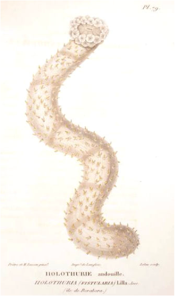
Fig. Original drawing of H. hilla; remark the very clearly drawn transversal banding, the many tube feet and the coloration pattern.
For original description click here.
{morfeo 44}